
PUMPA - SMART LEARNING
எங்கள் ஆசிரியர்களுடன் 1-ஆன்-1 ஆலோசனை நேரத்தைப் பெறுங்கள். டாப்பர் ஆவதற்கு நாங்கள் பயிற்சி அளிப்போம்
Book Free Demo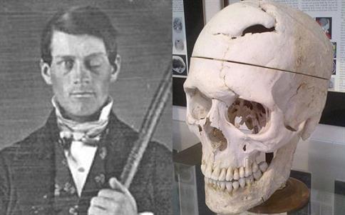
Phineas Gage and his skull after the accident
Unfortunately, the powder exploded, sending the one-metre-long, three-centimetre-diameter rod soaring upward. Before landing, the rod pierced Gage's left cheek, tore into his brain, and exited his skull.
Gage amazingly survived the accident. Due to the damage to his left frontal lobe of the brain, his personality and behaviour, on the other hand, were so drastically altered and, as a result, that many of his friends described him as a completely new person. This was a shocking revelation for experts. For the first time, there was proof that brain damage could influence our behaviour and personality.
This incident changed the course of neuroscience, and several brain specialists used information from Phineas case to support their own views about how the brain operates.
Regions of the brain
Within each of us lies one of the most fascinating and wondrous things in the universe is our brain. Our brain is remarkably little for everything it does, weighing only around 1.5 kg and containing 100 billion neurons that allow us to feel, see, hear, smell, move, think, laugh, cry, speak, read, and remember. The anatomy of our brain is unique, with various regions and folds that offer information.
Important!
Brain is the control and coordinating centre of the body. It controls the entire processing of information and act as a central information processing organ of our body.
Also, it controls and regulates,
- Voluntary movements of the body
- Balance of the body
- Function of involuntary organs such as lungs, heart, kidneys etc.
- Temperature of the body
- Endocrine glands
Meninges of the brain
Brain is the site for processing of vision, hearing, speech, memory, intelligence, emotions and thoughts. Human brain is protected by skull. Inside the skull, brain is surrounded by cranial meninges. It consists of three layers viz.,
- Dura mater - outer layer
- Arachnoid mater - middle layer
- Pia mater - inner layer
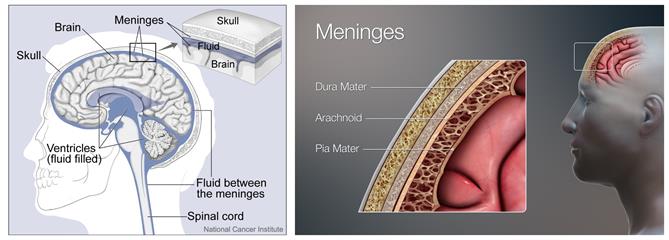
The location of cranial meninges and cerebrospinal fluid
Brain, being the largest part of the central nervous system, has three general areas.
- Brain stem
- Cerebral cortex
- Cerebellum
In healthy people, these three areas work together flawlessly, allowing the brain to coordinate critical activities and behaviours such as breathing and spatial navigation.
Parts of the brain
There are three major parts or regions of the brain viz.,
- Forebrain
- Midbrain
- Hindbrain
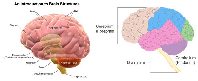
Pictures showing the regions of the brain
I. Forebrain
- Forebrain is the region mainly responsible for the process of thinking.
- It has regions that receive sensory impulses through various receptors.
- Specialised areas of the forebrain are responsible for hearing, smelling, sight and so on.
- There are specific areas of association where the sensory information received through hearing, smelling, and sighting is integrated with information from other receptors and information stored in the brain in the form of memory.
- Based on the integration of all this information, a decision is taken about how to respond. Then this decision will be sent to the motor area, which is the controlling region for the movement of voluntary muscle.
- Apart from the sensations of hearing, smelling, and sight, sensations such as feeling full happen during eating. This sensation is due to the specialised sensory region in the forebrain, which is associated with hunger.
Forebrain consists of,
- Olfactory lobes
- Cerebrum
- Diencephalon
1. Olfactory lobes: They are paired club shaped structures present in the anterior part of the brain. These lobes are responsible for the sense of smell.
2. Cerebrum:
It is the largest part of the brain. Cerebral cortex forms the outermost portion of cerebrum and makes up the grey matter.
Cerebral cortex, which sits above brainstem and cerebellum, swiftly perceives, interprets, and responds to information from the environment. Sensory perception and processing, as well as higher-level cognitive tasks including perception, memory, and decision-making, are all handled by it. \
Cerebral cortex is divided into two hemispheres and Corpus callosum, a bridge of large, flat neural fibres acts as a communication relay between the two sides.
Each cerebral hemisphere consists of four lobes namely,
- Frontal Lobe
- Parietal Lobe
- Occipital Lobe
- Temporal Lobe
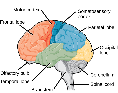
The lobes of the brain
- Frontal lobe: From behind the forehead back to the parietal lobe, the big frontal lobe stretches. It is the command centre for executive functions such as reasoning, decision-making, expressive language, higher-level cognitive processes, orienting (sensory information integration into person, place, time, and circumstance), and movement planning and execution.
- Parietal lobe: Above the temporal lobe and adjacent to the occipital lobe lies the parietal lobe. This lobe plays an important role in touch and spatial navigation, including the processing of touch, pressure, temperature, and pain.
- Occipital Lobe: The occipital lobe is present at the back of the brain. This region is responsible for processing and interpreting visual information.
- Temporal lobe: Temporal lobe is present from the temple back towards the occipital lobe. This region act as processing center for receptive language, memory and emotion.
The inner part of cerebral hemispheres and the structures like amygdala, hippocampus form a complex structure called the limbic system. Along with hypothalamus, this system regularizes the sexual behaviour and expression of emotional reactions and motivation.
3. Diencephalon:
It is consisting of,
- Epithalamus
- Thalamus
- Hypothalamus
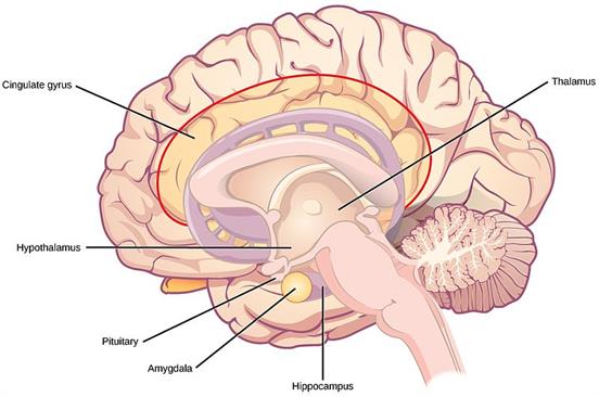
The regions of thalamus and hypothalamus
- Epithalamus secretes the hormone melatonin.
- Thalamus lies superior to midbrain. It is mainly composed of grey matter. Except smell, each sense channels and their sensory nerves pass through the thalamus.
- Hypothalamus lies above pituitary gland and is attached to it by a stalk called infundibulum. This region maintains homeostasis, controls the urge for eating and drinking. Neuro secretory cells in this region secrete hormone called as hypothalamic hormones.
II. Midbrain
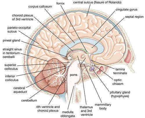
Location: Between the thalamus/hypothalamus of the forebrain and pons of the hindbrain.
Cerebral aqueduct, a canal, passed through the midbrain.
Midbrain is composed of corpora quadrigemina and crura cerebri.
- Corpora quadrigemina has two parts: (i) Superior colliculi - sense of sight (ii) Inferior colliculi - hearing
- Crura cerebri relay impulses back and forth between the cerebrum, cerebellum, pons and medulla.
III. Hindbrain
Hindbrain act as control centres for many of the involuntary actions, and it consists of following parts,
- Pons or Pons varolii present below the midbrain consists of fibre tracts that interconnect different regions of the brain. This region regulates the
- Cerebellum consists of highly convoluted surface to provide additional space for more neurons. It is responsible for maintaining the posture and balance of the body while doing voluntary actions such as walking on a narrow column, riding a bicycle, picking up a pen. The cerebellum also controls the precision of these voluntary actions.
- Medulla also called as medulla oblongata contains control centres of respiration, cardiovascular reflexes and gastric secretions. It is connected to spinal cord. Salivation, maintaining of blood pressure and vomiting are some of the involuntary actions controlled by the medulla in the hindbrain.
Important!
Note:
When individuals drink alcohol, it affects the cerebellum, which causes changes in movement and balance. This is the reason that drunk persons are more prone to collapse or have impaired speech.
Brainstem
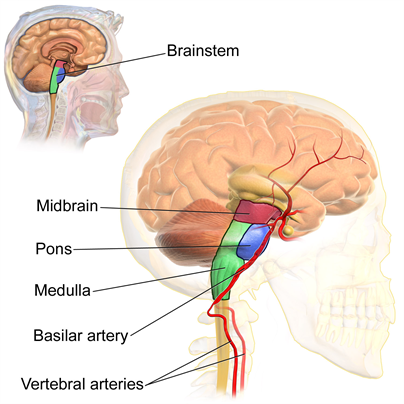
Parts of the brain stem
It connects the brain to the spinal cord. It is responsible for autonomic control of activities such as respiration and heart rate, as well as information transmission to and from the peripheral nervous system, which includes nerves and ganglia outside the brain and spinal cord. It is the place of entry or exit for ten of twelve cranial nerves. It is composed of,
- Diencephalon
- Medulla oblongata
- Pons varolii
- Mid brain
Grey matter and white matter:
-
The cell bodies and dendrites of neurons, as well as supporting cells called astroglia and oligodendrocytes, make up grey matter.
-
White matter, on the other hand, is largely made up of axons that are wrapped in myelin.
-
Grey matter neurons do not have long axons, unlike white matter neurons.
-
Grey matter accounts for 40% of the brain's volume, whereas white matter accounts for 60%.
-
Grey matter gets its colour from the grey nuclei that constitute the cells. The white appearance of the white matter is due to myelin.
-
Grey matter completes processing, whereas white matter facilitates communication between grey matter regions and between grey matter and other parts of the body.
-
Grey matter does not have a myelin coating, but white matter possesses it.
Important!
Myelin is an insulating substance that helps cells transport messages faster, and it is responsible for the white matter's lighter colour.
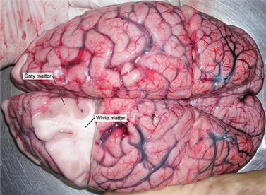
Grey and white matter in the brain
Spinal cord:
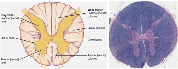
The cross-sectional view of the spinal cord with grey and white matter
Spinal cord extends from the medulla oblongata for about 42 to 45 cm long. It acts as means of communication between spinal nerves and brain. It carries impulses to and from the brain to the controls most of the reflex actions.
Reference:
https://upload.wikimedia.org/wikipedia/commons/8/83/Diagram_showing_the_brain_stem_which_includes_the_medulla_oblongata%2C_the_pons_and_the_midbrain_%282%29_CRUK_294.svg
https://upload.wikimedia.org/wikipedia/commons/e/e8/Figure_35_03_03.png
https://commons.wikimedia.org/wiki/File:Blausen_0115_BrainStructures.png
https://upload.wikimedia.org/wikipedia/commons/e/e0/Blausen_0114_BrainstemAnatomy.png
https://www.britannica.com/science/medulla-oblongata
https://upload.wikimedia.org/wikipedia/commons/1/16/1313_Spinal_Cord_Cross_Section.jpg
https://www.flickr.com/photos/nihgov/26276988755
https://upload.wikimedia.org/wikipedia/commons/1/13/3D_Medical_Illustration_Meninges_Details.jpg
https://upload.wikimedia.org/wikipedia/commons/thumb/a/af/1202_White_and_Gray_Matter.jpg/512px-1202_White_and_Gray_Matter.jpg
https://upload.wikimedia.org/wikipedia/commons/thumb/b/b6/Corpus_callosum.jpg/512px-Corpus_callosum.jpg