
PDF chapter test TRY NOW
Based on the shape of the cells and their arrangement, epithelial tissues further classified as follows:
- Covering epithelial tissue
- Glandular epithelial tissue
I. Covering epithelial tissue
Covering epithelial tissue located over the surface of the body parts, both externally and internally. Based on the number of cell layers, it is classified into,
- Simple epithelial tissue
- Stratified epithelial tissue
1. Simple epithelial tissue
Simple epithelial tissue consists of a single layer of cells, which is resting on the basement membrane. It is further classified into,
- Simple squamous epithelial tissue
- Simple cuboidal epithelial tissue
- Simple columnar epithelial tissue
- Simple ciliated epithelial tissue
- Pseudostratified epithelial tissue
Based on the shape of cells on the exposed surface, squamous (tile-like), cuboidal and columnar are classified.
A. Simple squamous epithelial tissue
- It is a single-layered epithelium.
- The cells are very thin and flat, with disc-shaped central nuclei and sparse cytoplasm with irregular boundaries.
- It is also called pavement membrane due to its tile-like appearance.
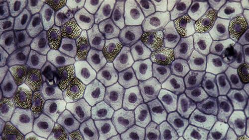
Simple squamous epithelial tissue
Location: Simple squamous epithelia are located in the lungs, air sacs, lining of the heart, kidney glomeruli, blood vessels, and lymph vessels. It also covers the lining of the oesophagus and the lining of the mouth.
Functions:
- It forms a delicate lining of blood vessels in lung alveoli, where substance transport occurs across a selectively permeable membrane by diffusion.
- Secretes lubricating substance.
- It helps in filtration by forming a selectively permeable membrane surface.
B. Simple cuboidal epithelial tissue
- The height and width of the cells almost look equal, i.e., cube-shaped cells.
- The nucleus is round in shape and located at the centre of the cell.
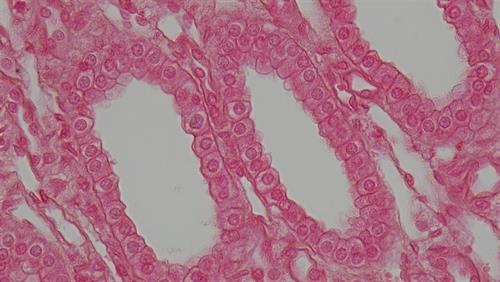
Simple cuboidal epithelial tissue
Location: Epithelia of kidney tubules and many glands.
Functions:
- It helps in the absorption of useful substance from urine.
- Secretion of gastric juices.
C. Simple columnar epithelial tissue
It is made up of tall, cylindrical pillar-like cells with oval nuclei, facilitating movement across the epithelial barrier.
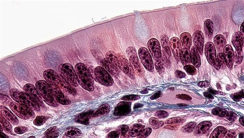
Simple columnar epithelial tissue
Location: It is usually found in the stomach and intestine's inner lining, where absorption and secretion occurs.
Function: Absorption of nutrients from the digested food, secretion of mucus and enzymes.
D. Simple ciliated epithelial tissue
It consists of cilia, i.e., hair-like projections, which can move due to the fine, vibratile cytoplasmic processes.
It is of two types,
- Cuboidal ciliated epithelium
- Columnar ciliated epithelium
a. Simple cuboidal ciliated epithelium:
Location: Certain parts of the kidney's urinary tubules.
Function: Helps in urine's movement.
b. Simple columnar ciliated epithelium:
Location: Linings of nasal passages, oviducts, terminal bronchioles, ventricles of the brain.
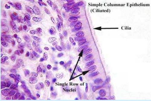
Simple columnar ciliated epithelium
Function: Their movement pushes substances like mucus forward to clear it from the ducts. The beating of the cilia moves solid dust particles in one direction through ducts.
E. Pseudostratified epithelial tissue
It is a single layer epithelium but appears as stratified due to the uneven length of the cells.
It is of two types,
a. Pseudostratified columnar epithelial tissue
b. Pseudostratified columnar ciliated epithelial tissue
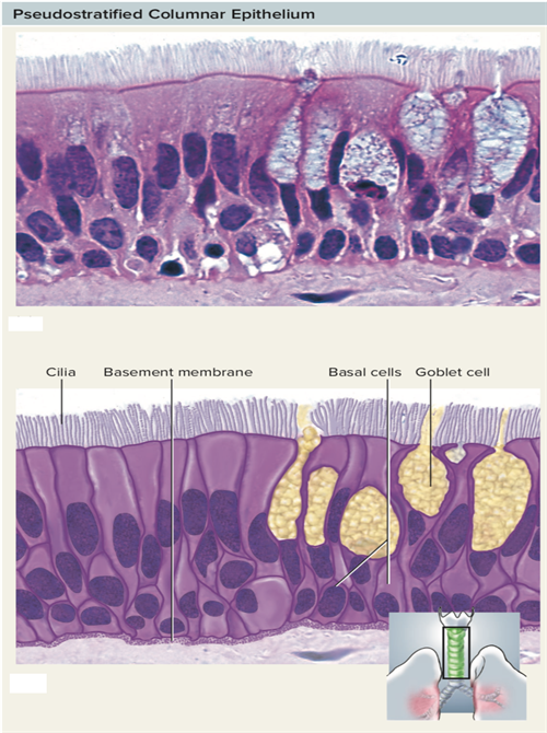
Pseudostratified columnar epithelial tissue
a. Pseudostratified columnar epithelial tissue:
Location: Ducts of parotid salivary gland, olfactory mucosa
Function: Absorption and filtration processes.
b. Pseudostratified columnar ciliated epithelial tissue:
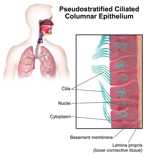
Pseudostratified columnar ciliated epithelial tissue
Location: Linings of trachea and bronchi.
Function: Secretes mucous, ciliated tissue move mucous.
Reference:
https://upload.wikimedia.org/wikipedia/commons/e/e6/Epithelial_Tissues_Simple_Squamous_Epithelium_%28frog%29_%2827847646938%29.jpg
https://upload.wikimedia.org/wikipedia/commons/9/9e/Epithelial_Tissues_Simple_Cuboidal_Epithelium_%2841681552782%29.jpg
https://commons.wikimedia.org/wiki/File:Epithelial_Tissues_Brush_Border_in_Simple_Columnar_Epithelium_(27854453998).jpg
https://upload.wikimedia.org/wikipedia/commons/1/16/Ciliated_columnar_epithelium.JPEG
https://commons.wikimedia.org/wiki/File:2304_Pseudostratified_Epithelium.jpg
https://upload.wikimedia.org/wikipedia/commons/3/33/Blausen_0750_PseudostratifiedCiliatedColumnar.png
 Register for free to see more content