
PUMPA - SMART LEARNING
எங்கள் ஆசிரியர்களுடன் 1-ஆன்-1 ஆலோசனை நேரத்தைப் பெறுங்கள். டாப்பர் ஆவதற்கு நாங்கள் பயிற்சி அளிப்போம்
Book Free Demo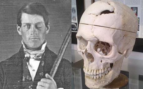
Phineas Gage and his skull after the accident
Gage, then 25 years old, was the foreman of a group preparing a railroad bed in Cavendish, Vermont, on September 13, 1848. He was tamping explosive material into a hole with an iron tamping rod.
Unfortunately, the powder exploded, sending the one-metre-long, three-centimetre-diameter rod soaring upward. Before landing, the rod pierced Gage's left cheek, tore into his brain, and exited his skull.
Gage amazingly survived the accident. Due to the damage to his left frontal lobe of the brain, his personality and behaviour drastically altered and many of his friends described him as a completely new person. This was a shocking revelation for experts. For the first time, there was proof that brain damage could influence our behaviour and personality.
This incident changed the course of neuroscience, and several brain specialists used information from Phineas case to support their views about how the brain operates.
Regions of the brain:
One of the most fascinating and wondrous things in the universe within each of us is our brain. Our brain is remarkably little for everything it does, weighing only around 1.5 kg and containing 100 billion neurons that allow us to feel, see, hear, smell, move, think, laugh, cry, speak, read, and remember.
The anatomy of our brain is unique, with various regions and folds that offer information.
Important!
The brain is the control and coordinating centre of the body. It controls the entire processing of information and acts as a central information processing organ of our body.
Also, it controls and regulates,
- Voluntary movements of the body
- Balance of the body
- Functions of involuntary organs such as lungs, heart, kidneys, etc.
- Temperature of the body
- Endocrine glands
Meninges of the brain:
Brain is the site for processing vision, hearing, speech, memory, intelligence, emotions and thoughts. The human brain is protected by the skull. Inside the skull, the brain is surrounded by three connective tissue membranes or cranial meninges. Besides the skull, it also protects the brain from mechanical injury. The three membranes of the meninges are,
- Dura mater
- Arachnoid membrane
- Pia mater
Type of membrane | Origin of the name | Location | Structure |
| Dura mater | dura: tough; mater: membrane | outermost layer | Thick fibrous membrane |
| Arachnoid membrane | arachnoid: spider | middle layer | Thin vascular membrane providing web-like cushion |
| Pia mater | Pia: soft or tender | innermost layer | Thin delicate membrane richly supplied with blood |
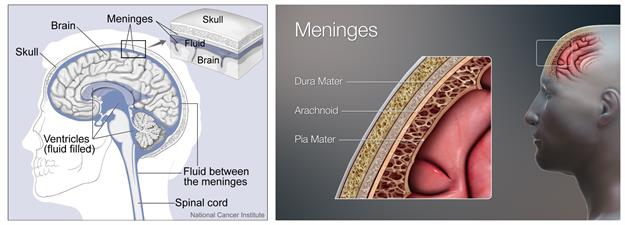
The location of cranial meninges and cerebrospinal fluid
Important!
Meningitis is a disease caused due to the inflammation of the meninges. When the fluid around the meninges becomes infected, Meningitis might occur. Infections caused by viruses and bacteria are the most common causes of meningitis.
Also, there is a clear fluid called Cerebrospinal fluid (CSF) present in ventricles of the brain, subarachnoid space of the brain and spinal cord. It acts as a shock-absorbing medium.
The brain, being the largest part of the central nervous system, has three general areas.
- Brain stem
- Cerebral cortex
- Cerebellum
In healthy people, these three areas work together flawlessly, allowing the brain to coordinate critical activities and behaviours such as breathing and spatial navigation.
Parts of the brain:
There are three major parts or regions of the brain viz.,
- Forebrain
- Midbrain
- Hindbrain
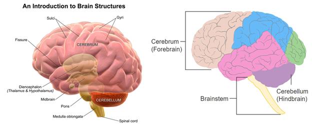
Pictures showing the regions of the brain
I. Forebrain:
The Forebrain consists of,
- Olfactory lobes
- Cerebrum
- Diencephalon
1. Olfactory lobes: They are paired club-shaped structures present in the anterior part of the brain. These lobes are responsible for the sense of smell.
2. Cerebrum:
It is the largest part of the brain and occupies nearly two-thirds of the brain. The cerebral cortex forms the outermost portion of the cerebrum and makes up the grey matter.
The innermost or deeper portion, made of white matter, is called the cerebral medulla. The cortex is heavily folded, forming elevations called gyri and depressions between them termed sulci, increasing its surface area.
The cerebral cortex, which sits above the brainstem and cerebellum, swiftly perceives, interprets, and responds to information from the environment. Sensory perception and processing, as well as higher-level cognitive tasks including perception, memory, and decision-making, are all handled by it.
Cerebrum is divided longitudinally into two hemispheres (right and left hemispheres) by a deep cleft called as median cleft. Corpus callosum, a bridge of large, flat, thickband of neural fibres, acts as a communication relay between the two sides.
Each cerebral hemisphere consists of four lobes called as cerebral lobes viz.,
- Frontal Lobe
- Parietal Lobe
- Occipital Lobe
- Temporal Lobe
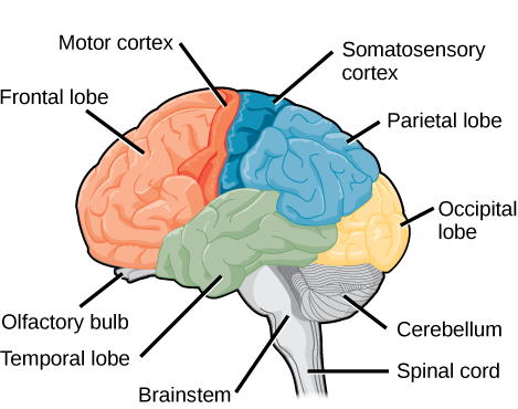
The lobes of the brain
i. Frontal lobe:
From behind the forehead back to the parietal lobe, the big frontal lobe stretches. It is the command centre for executive functions such as reasoning, decision-making, expressive language, higher-level cognitive processes, orienting (sensory information integration into person, place, time, and circumstance), and movement planning and execution.
ii. Parietal lobe:
Above the temporal lobe and adjacent to the occipital lobe lies the parietal lobe. This lobe plays an important role in touch and spatial navigation, including the processing of touch, pressure, temperature, and pain.
iii. Occipital Lobe:
The occipital lobe is present at the back of the brain. This region is responsible for processing and interpreting visual information.
iv. Temporal lobe:
The temporal lobe is present from the temple back towards the occipital lobe. This region act as the processing center for receptive language, memory and emotion.
Important!
The inner part of the cerebral hemispheres and the structures like the amygdala, hippocampus form a complex structure called the limbic system. Along with the hypothalamus, this system regularises sexual behaviour and the expression of emotional reactions and motivation.
3. Diencephalon:
It is consists of,
- Epithalamus
- Thalamus
- Hypothalamus
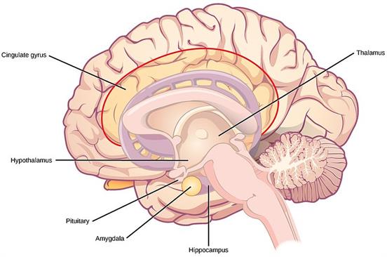
The regions of the thalamus and hypothalamus
Epithalamus: It secretes the hormone melatonin.
Thalamus: It lies in the cerebral medulla region. It is mainly composed of grey matter. It acts as a major conducting centre for the sensory and motor signalling. So, it is called relay centre.
Important!
Note: Except smell, each sense channels and their sensory nerves pass through the thalamus.
Hypothalamus: It lies above the pituitary gland and at the base of the thalamus. It is attached to the pituitary gland by a stalk called the infundibulum.
- This region maintains homeostasis and involves involuntary functions like hunger, thirst, sleep, sweating, sexual desire, anger, fear, water balance, blood pressure, etc.
- The body temperature is regulated in this region, so it is called the thermo-regulatory centre of the body.
- Neuro secretory cells in this region secrete a hormone called hypothalamic hormones. These hormones control anterior pituitary's hormone production and secretion.
- The hypothalamus is an anatomical link between the nervous and endocrine systems. It is regarded as the endocrine system's supreme commander.
Reference:
https://upload.wikimedia.org/wikipedia/commons/8/83/Diagram_showing_the_brain_stem_which_includes_the_medulla_oblongata%2C_the_pons_and_the_midbrain_%282%29_CRUK_294.svg
https://upload.wikimedia.org/wikipedia/commons/e/e8/Figure_35_03_03.png
https://commons.wikimedia.org/wiki/File:Blausen_0115_BrainStructures.png
https://www.britannica.com/science/medulla-oblongata
https://www.flickr.com/photos/nihgov/26276988755
https://upload.wikimedia.org/wikipedia/commons/1/13/3D_Medical_Illustration_Meninges_Details.jpg
https://upload.wikimedia.org/wikipedia/commons/thumb/b/b6/Corpus_callosum.jpg/512px-Corpus_callosum.jpg