
PUMPA - SMART LEARNING
எங்கள் ஆசிரியர்களுடன் 1-ஆன்-1 ஆலோசனை நேரத்தைப் பெறுங்கள். டாப்பர் ஆவதற்கு நாங்கள் பயிற்சி அளிப்போம்
Book Free DemoTissue system and its functions:
Tissue system | Components | Functions |
| Dermal tissue system | Epidermis and periderm (In older stem and roots) |
|
| Ground tissue system | Parenchyma tissue Collenchyma tissue Sclerenchyma tissue |
|
| Vascular tissue system | Vascular tissue Xylem tissue Phloem tissue |
|
Differences between dicot and monocot root:
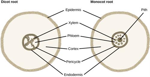
Dicot and monocot root
Tissues | Dicot root | Monocot root |
| Number of xylem | Tetrarch | Polyarch |
| Cambium | Present(During secondary growth only) | Absent |
| Secondary growth | Present | Absent |
| Pith | Absent | Present |
| Conjunctive tissue | Parenchyma | Sclerenchyma |
| Example | Bean | Maize |
Differences between dicot stem and monocot stem:
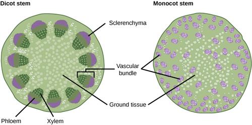
Dicot and monocot stem
| Tissues | Dicot stem | Monocot stem |
| Hypodermis | Collenchymatous | Sclerenmatous |
| Ground tissue | Differentiated into cortex, endodermis, pericycle and pith | Undifferentiated |
| Vascular bundles |
|
|
| Secondary growth | Present | Mostly absent |
| Pith | Present | Absent |
| Medullary rays | Present | Absent |
Differences between of dicot and monocot leaf:
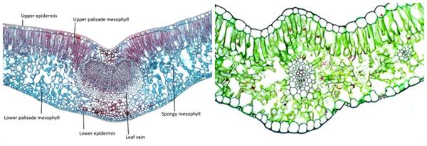
Left to right: Dicot and monocot leaf
Dicot leaf | Monocot leaf |
Dorsiventral leaf | Isobilateral leaf |
Mesophyll is differentiated into palisade and spongy parenchyma | Mesophyll is not differentiated into palisade and spongy parenchyma |
Reference:
https://commons.wikimedia.org/wiki/File:Figure_30_03_04.jpg
https://www.flickr.com/photos/71183136@N08/6985212944
https://en.m.wikipedia.org/wiki/File:Dicot_leaf_L.jpg
https://commons.wikimedia.org/wiki/File:Figure_30_02_06.jpg
 Register for free to see more content