PUMPA - SMART LEARNING
எங்கள் ஆசிரியர்களுடன் 1-ஆன்-1 ஆலோசனை நேரத்தைப் பெறுங்கள். டாப்பர் ஆவதற்கு நாங்கள் பயிற்சி அளிப்போம்
Book Free Demo
Monocot stem
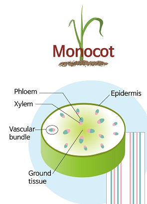
In monocot stem, vascular bundles are closed due to the absence of cambium. So they don't have secondary growth, and tree rings are also absent in it. The internal structure of the monocot stem is explained in this topic.

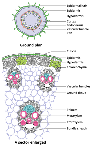
Transverse section of monocot stem
The above picture is showing a sector of the transverse section of a monocot stem.
The following structures are seen in the section.
i. Epidermis:
It is the single outermost layer present in the monocot stem. It is made up of parenchyma cells. Cuticle, a thick protective layer, covers the outer wall of this layer. It aids in the prevention of water evaporation due to transpiration. Multicellular hairs are absent, and stomata are also less in number.
ii. Hypodermis:
This region is made up of sclerenchyma cells interrupted by chlorenchyma cells. Sclerenchymatous cells provides mechanical support to the plant.iii. Ground tissue:
An area below the hypodermis is made up of a whole mass of parenchyma cells with intercellular spaces extending to the centre.This area is referred to as ground tissue. No differentiation of endodermis, cortex, pericycle and pith is observed in the monocot stem.iv. Vascular Bundle:
a. Xylem:
Both protoxylem and metaxylem are present in the vascular bundle. These xylem vessels are arranged in V or Y shape. In the mature vascular bundle, the protoxylem present at the lowermost point disintegrates and form a cavity. This cavity is called protoxylem lacuna.
Both protoxylem and metaxylem are present in the vascular bundle. These xylem vessels are arranged in V or Y shape. In the mature vascular bundle, the protoxylem present at the lowermost point disintegrates and form a cavity. This cavity is called protoxylem lacuna.
b. Phloem:
Phloem components - Sieve tube elements and companion cells are present. However, Phloem parenchyma and phloem fibers are absent.
v. Pith:
Pith is not differentiated in monocot stems.
Video explaining the internal structure of monocot stem