
PUMPA - SMART LEARNING
எங்கள் ஆசிரியர்களுடன் 1-ஆன்-1 ஆலோசனை நேரத்தைப் பெறுங்கள். டாப்பர் ஆவதற்கு நாங்கள் பயிற்சி அளிப்போம்
Book Free DemoIn the vertebrate body, supportive or skeletal connective tissue forms the endoskeleton. These tissues support and protect the organs and also help in locomotion. Cartilage and bone are supporting tissues.
If we fold the ears and nose, they can bend easily due to cartilage, but we cannot bend the bones present in the arms and legs. Cartilage is mainly present in the embryonic stages and acts as a supporting skeleton. Bones replace a majority of the cartilage in adults.
Cartilage:
It is a connective tissue having widely spaced cells. It is a non-porous, semi-rigid, flexible tissue. It has a solid matrix called chondrin which is made up of proteins and sugars. The matrix contains large cartilage cells called chondrocytes. These cells are found in fluid-filled spaces known as lacunae.
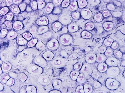
Microscope view of human cartilage
Location : In humans, cartilage is present in the tip of the nose, external ear, end of long bones, trachea and larynx.
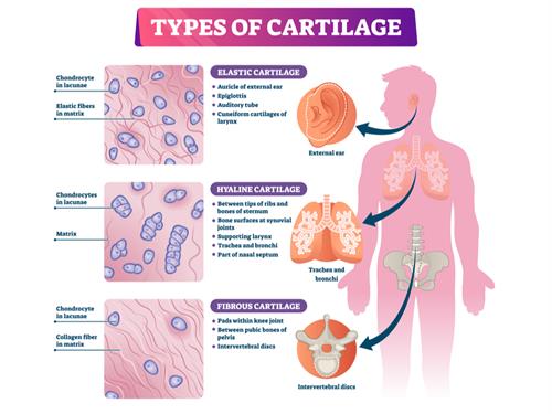
Different types of cartilage present in the human body
Function:
- Cartilage gives smoothness to the bone surfaces where it is joined.
- Its principal function is to connect bones.
- Cartilage provides support, act as a shock absorber, and provide a smooth, flexible, friction-free surface that allows our joints to work and our bones to move against each other painlessly.
Bone:
If a person has a fracture in his leg, how could he facilitate himself for the movement? He may use crutches for the movement. Crutches gives mechanical support for him to walk. What gives you the mechanical support for the movement, just like crutches, when you do not need them? The answer is bones.
Bones provide mechanical support for our body as an endoskeleton. It gives shape and a structural framework to the body.
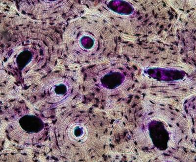
Compact bone - ground cross section
Bone is a solid, rigid, and strong non-flexible hard connective tissue that forms vertebrates' skeleton. It is porous, highly vascular(Comprises blood vessels and cells), mineralized, stiff and rigid. The cells are embedded inside the hard matrix, which is made up of proteins, collagen fibres, and rich in salts of calcium, phosphorous, which are the main reasons for the bone'shardness and strength.
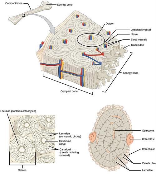
Internal structure of bone
Important!
The bone matrix is in the form of bright and dark concentric rings called lamellae.
The fluid-filled spaces present between the lamellae is called lacunae, in which the bone cells are called osteocytes.
Each bone cells communicate with one another by a network of fine canals called canaliculi.
The hollow cavities of spaces are called marrow cavities filled with bone marrow.
The fluid-filled spaces present between the lamellae is called lacunae, in which the bone cells are called osteocytes.
Each bone cells communicate with one another by a network of fine canals called canaliculi.
The hollow cavities of spaces are called marrow cavities filled with bone marrow.
Function:
- Bones provides shape and structural framework to the body.
- Bones protect the vital organs such as the brain, lungs from injury.
- It also anchors the muscles and serves as a storage site of calcium and phosphate.
- Bones enable body movements by acting as a push button and points of attachment for muscles.
- The formation of blood cells (hematopoiesis) occurs in the red marrow found within the cavities of certain bones.
- Bones serve as an energy reservoir by storing lipids in the form of fats in adipose tissues.
Types of bone cells:
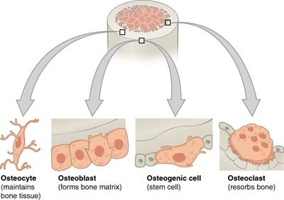
- The process of bone formation is termed as osteogenesis.
- Osteoblast cells are responsible for the new bone formation.
Reference:
https://upload.wikimedia.org/wikipedia/commons/0/0c/Compact_bone_-_ground_cross_section.jpg
https://commons.wikimedia.org/wiki/File:604_Bone_cells.jpg
https://upload.wikimedia.org/wikipedia/commons/4/49/Figure_33_02_09.jpg
https://commons.wikimedia.org/wiki/File:604_Bone_cells.jpg
https://upload.wikimedia.org/wikipedia/commons/4/49/Figure_33_02_09.jpg