
PUMPA - SMART LEARNING
எங்கள் ஆசிரியர்களுடன் 1-ஆன்-1 ஆலோசனை நேரத்தைப் பெறுங்கள். டாப்பர் ஆவதற்கு நாங்கள் பயிற்சி அளிப்போம்
Book Free Demo“I think; therefore, I am."
This is a famous statement made by the French philosopher Descartes. The process of thinking made him realise that he existed. Thinking makes us who we are. Thinking and reasoning are such important things which gives all the advances for the humankind.
It is done by a special type of tissue called nervous tissues or neural tissues. Besides thinking and reasoning, it coordinates and controls many body activities. It also functions to stimulate muscle contraction, create awareness of the environment, and create emotions and memory.
Neural tissue develops from the ectoderm of the embryo, except microglia, which originate from the mesoderm. It is found throughout the body. A neural tissue consists of neurons and neuroglia.
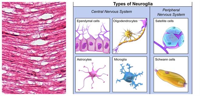
Microscopic view of nervous tissue and types of neuroglia
The unique properties of the cells of the neural tissues are excitability and conductivity. These are highly specialized for receiving stimulus and then transmitting it very rapidly from one place to another within the body itself.
Location: Nervous tissue abundantly occurs in the brain, spinal cord, and nerves. The tissue forms the nervous system, consisting of three parts,
- Central nervous system (CNS)
- Peripheral nervous system (PNS)
- Autonomic nervous system (ANS)
Structural and functional units of neural tissue are called neurons or nerve cells.
Neurons are the body's longest cells. A single neuron can be up to a metre long and is made up of three main components:
- Cell body
- Axon
- Dendrites
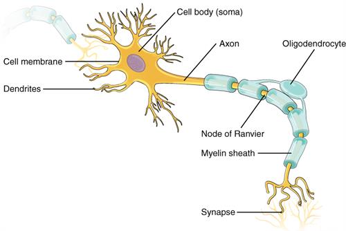
Structure of a neuron
1. Cell body (Cyton):
- It is located in the grey matter of the brain's central nervous system, but a few are found outside the CNS and are known as ganglia.
- Cytoplasm, nucleus, and cell organelles such as mitochondria, golgi bodies, endoplasmic reticulum, ribosomes, and lysosomes make up the cell.
- The centrosome is absent in neurons.
Neuritis is a term used to describe two types of neuron processes. Axon and dendrites (or dendrons). The number of dendrites varies, but the number of axons is always one.
2. Axon: It is a single, long, cylindrical conducting fibre extending from a neuron.
Function: It transmits impulse away from the cell body.
3. Dendrites: These are short branched, hair-like fibres of neuron. Nissl bodies, neurofibrils and mitochondria are present in dendrites.
Function: It carry information from their tips towards the axon.
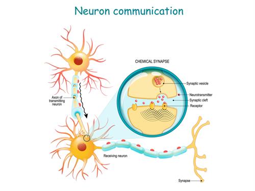
Neurons communicate with each other by imparting neurotransmitters via contact points, the so-called neuronal synapses.
Note:
- All neurons are distinguished by the presence of Nissl bodies and neurofibrils. Nissl bodies are ribosome and rough endoplasmic reticulum masses that are relatively wide.
- Synapse is a region of union of one neuron's axon with the dendrite of the next neuron. It allows the transfer of nerve impulse generated to and from in the body.
- Information flow in a neuron is unidirectional. Dendrites receive signals from other neurons via specialized interfaces called synapses. After that, axons transmit the signal to the next cell, often over long distances.
- Many nerve fibres joined together by connective tissue and formed a nerve.
- Nerve and muscle tissues are the fundamental units of animals, and this combination enables animals to move rapidly in response to stimuli.
- Chemicals used for neurotransmission are called neurotransmitters, e.g. Dopamine, Serotonin and Acetylcholine.
There are four groups of neurons based on their functional existence to structure.
- A polar neurons
- Unipolar neurons
- Bipolar neurons
- Multipolar neuron
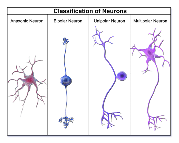
Types of neuron
1. A polar neurons:
- They don't have any polarity.
- Axons and dendrites are not distinguished in these fibres.
- All fibres are of the same kind and can carry information towards or away from the cell body.
Example:
Neurons of a hydra.
2. Unipolar neurons:
- They have one axon and one dendrite only.
- The flow of information is unidirectional.
- These are sensory neurons.
Example:
They can be found in invertebrate and vertebrate embryos.
3. Bipolar neurons: With one dendron and one axon at opposite poles, these neurons have a unidirectional information flow.
Example:
They can be found in the retina of the eyes, as well as the olfactory epithelium.
4. Multipolar neuron:There are neurons with a single axon and several dendrites that provide a unidirectional information flow.
Example:
They can be found in the nervous system of adult vertebrates.
Based on function, neurons are of three types.
- Sensory neurons
- Motor neurons
- Relaying neurons
1. Sensory neurons/Afferent neurons: Sensory neurons transmit nerve impulses by connecting sensory or receptor cells or organs to the central nervous system.
2. Motor/efferent neurons:Motor neurons connect CNS to muscle and glands.
3. Relaying neurons: They occur between the sensory and motor neurons in the brain and spinal cord for the distant transmission of nerve impulses.
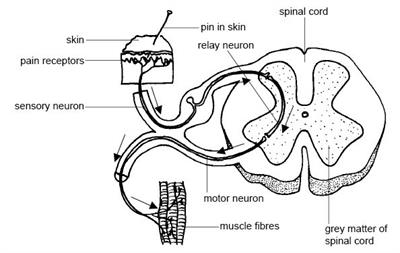
Anatomy and physiology of animals Relation btw sensory, relay & motor neurons
Important!
Do you know!
Reference:
https://upload.wikimedia.org/wikipedia/commons/8/86/1206_The_Neuron.jpg
https://commons.wikimedia.org/wiki/File:Nervous_Tissue_Nerve_Bundle_(27048520767).jpg
https://commons.wikimedia.org/wiki/File:Neuron_Classification.png
https://commons.wikimedia.org/wiki/File:Blausen_0870_TypesofNeuroglia.png
https://upload.wikimedia.org/wikipedia/commons/5/51/Anatomy_and_physiology_of_animals_Relation_btw_sensory%2C_relay_%26_motor_neurons.jpg