PUMPA - SMART LEARNING
எங்கள் ஆசிரியர்களுடன் 1-ஆன்-1 ஆலோசனை நேரத்தைப் பெறுங்கள். டாப்பர் ஆவதற்கு நாங்கள் பயிற்சி அளிப்போம்
Book Free DemoWe have studied, bones gives mechanical support for the movement. So, what part of our body helps us to move? Which part gets the signals from the brain, actually enables the movement in the body?
Muscular tissue is the answer.
Muscular tissues develop from the mesoderm of the embryo. Iris muscles of the eye develop from ectoderm. It is composed of muscle cells and forms the major part of contractile tissue. The muscle cells are elongated, and their size is large, and it contains several myofibrils.
Many long cylindrical fibrils makes the muscle, and they are arranged in parallel to one another. Muscle cells contain a special type of proteins called contractile proteins, which causes the body's movement by contraction and relaxation.
Based on the structure, location and function, the muscular tissues are further divided into three types:
- Skeletal muscles (or) Striated muscle
- Smooth muscle (or) Non-striated muscle
- Cardiac muscle
Skeletal muscles:
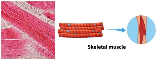
Skeletal muscles are closely attached to the skeletal bones and help in the body movement, e.g. limb (biceps and triceps of arms) muscles. Hence, these are called skeletal muscles.

Microscopic and schematic view of skeletal muscle
Location: It is found in the muscles of limbs, tongue, pharynx and beginning of the oesophagus.
- \(40\)% of our body mass made up of skeletal muscles.
- The muscle fibres are non- tapering, elongated, cylindrical, unbranched and multinucleate (having many nuclei).
- The muscles present in our limbs that we move or stop as per our will are skeleton muscles. These are also called voluntary muscles, as we can move them by conscious will.
- Each skeletal tissue contains myofibrils. A myofibril has dark and light bands.
- Under a microscope, striated muscles show alternate light and dark bands or striations. Thus, these are also known as striated muscles.
- The sarcomere is the fundamental contractile unit of muscle fibre.
- Light and dark bands of the sarcomere is made up of thick and thin protein filament such as myosin and actin, and these are the structures responsible for the rapid contraction.
Smooth muscles:
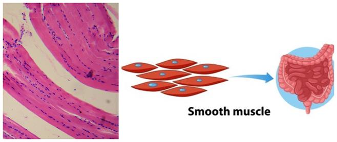
Location: It is found in the internal organs' walls, such as the blood vessels, gastric glands, intestinal villi, and urinary bladder.
The muscles which we cannot move as per our will are called smooth muscles. These are also called involuntary muscles.

Microscopic and schematic view of smooth muscle
- These cells are long muscles, spindle-shaped with a broad middle part, with pointed ends and uninucleate (single nucleus) located at the centre.
- These muscles do not show any dark or light band, So it is called as unstriated muscles.
Function: The movement of food in the alimentary canal or the contraction and relaxation of blood vessels are involuntary movements.
Cardiac muscles:
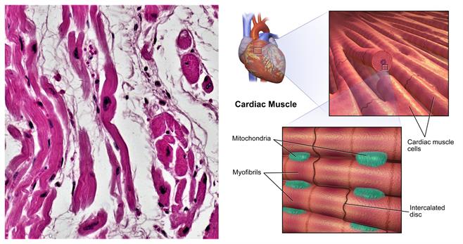
Location: It is a contractile tissue present only in the heart wall (Myocardium), having a very rich blood supply.
Cardiac muscles are involuntary muscles. They are constantly contracting and relaxing in a rhythmic pattern.

Microscopic and schematic view of cardiac muscle
- These muscles are involuntary, which means they are not beyond human control.
- The cells constituting cardiac muscles are cylindrical, uninucleate and branched.
- The cells of the cardiac muscles are cardiomyocytes, and it has stripes of light and dark bands.
- Cardiac cells are unique among the muscle types because they connect to form a network called intercalated discs.
- These cells have intercellular spaces, and they are filled with loose connective tissue supplied with blood capillaries.
Function: The human heart is myogenic, which means cardiac impulses arise from the Sinoatrial node (a node of specialized cardiac muscle fibres situated in the heart wall), resulting in rhythmic contraction and relaxation throughout the period it works.
Important!
The cytoplasm of a muscle fibre is termed as sarcoplasm.
Reference:
https://upload.wikimedia.org/wikipedia/commons/5/56/Skeletal_muscle_-_longitudinal_section.jpg
smooth muscle microscopic view
https://upload.wikimedia.org/wikipedia/commons/7/73/Cardiac_Muscle.png
https://upload.wikimedia.org/wikipedia/commons/2/2b/Muscle_Tissue_Cardiac_Muscle_%2828184528408%29.jpg
https://www.freepik.com/free-vector/type-muscle-cells_11573851.htm